

PROCEDURES


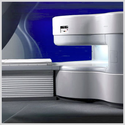
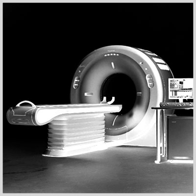
Open MRI
This state-of-the-art open MRI has a unique, "open on all 4 sides" design which is non-claustrophobic. It has a weight limit of 450 pounds. There are no loud knocking noises like closed-in magnets. Our MRI suite is custom designed and patient friendly. The MRI technologists are specially trained to perform your exam professionally while ensuring your maximum comfort. Click here for more details.
Click Here to Read more
Advanced Imaging Center's Open MRI features the Hitachi AIRIS Open MRI, the most advanced open MRI system. It was the first Open MRI in the Antelope Valley and still the only high-quality one. With our Open MRI you can enjoy the benefits of MRI without the fear of confinement.
This state-of-the-art open MRI has a unique, "open on all 4 sides" design which is non-claustrophobic. It has a weight limit of 450 pounds. There are no loud knocking noises like closed-in magnets. Our MRI suite is custom designed and patient friendly.
The MRI technologists are specially trained to perform your exam professionally while ensuring your maximum comfort. MRI, magnetic resonance imaging, is a method used by physicians to visualize internal organs of the human body and obtain diagnostic information. These images are produced without the use of radiation. MRI is a noninvasive procedure and there are no known side or after effects. The procedure is painless. You won't see or feel anything during the exam. A faint knocking sound will be heard which is the imaging process in operation.
MRI utilizes the physical properties of magnetic fields, radio waves, and computers to generate images of the body in any plane. The technique is an established, accurate diagnostic tool. It can quickly and noninvasively provide accurate diagnosis for your physician, which in some situations will reduce the need for exploratory surgery or other diagnostic procedures which may have greater risk.
The benefits of magnetic resonance imaging are vast and new applications are continually being developed through ongoing research. MRI imaging is used for virtually all parts of the body. It is the primary imaging modality for evaluation of diseases of the brain and spine. It is effective in depicting abnormalities of the eye, paranasal sinuses, throat, salivary glands, and the thyroid.
MRI is the method of choice for imaging of the musculoskeletal system and is widely used for evaluation of the shoulder, elbow, wrist, hip, knee, and ankle. It can also accurately depict abnormalities within the bone marrow.
It has many applications in the cardiovascular system. The display of blood vessels known as MR angiography is an accurate, noninvasive means of obtaining information about arteries of the head, neck, and body.
It is also effective in demonstrating congenital cardiac abnormalities. MRI is also important in evaluation of the organs of the abdomen and pelvis including the liver, pancreas, kidneys, ovaries, uterus, and prostate.


4D Ultrasound
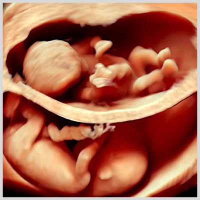
Advanced Imaging Center is proud to offer new revolutionary four-dimensional (4D) Ultrasound machines (award-winning GE Voluson 730 Expert scanners) with on-line real-time 4D capabilities. It is the same type featured on the Oprah television talk show and currently being used for example at Cedars Sinai Medical Center. Click here for more details.
Click Here to Read more
Advanced Imaging Center is proud to offer new revolutionary four-dimensional (4D) Ultrasound machines (award-winning GE Voluson 730 Expert scanners) with on-line real-time 4D capabilities. It is the same type featured on the Oprah television talk show and currently being used for example at Cedars Sinai Medical Center.
This amazing new technology is particularly useful in the OB patients for 4D visualization of the fetus (fetal face and other body parts, such as shown above). The mother will be given at least one 4D picture of the fetus at the end of the exam. Other applications include multiplanar imaging of the pelvic organs (including endovaginal 4D imaging). Other applications include multi-planar imaging (similar to MRI) of other body parts including abdomen, pelvis, small parts, neck, etc.
We have a full-time sonographer on the premises and offer a full range of ultrasound services including: abdominal, pelvic, OB, head & neck, small parts, breast, and vascular Doppler (carotid Duplex/CVP's, venous/DVT's, renal Doppler, peripheral arterial/venous Doppler, etc.).
A DVD with MUSIC is provided for the pregnant mom showing live 3D movie of her unborn baby.
Diagnostic ultrasound is an established method of diagnostic medical imaging using a high frequency sound wave and the principle of sonar. Because ultrasonic waves cause no damage to human tissues, they are an important tool utilized for both diagnosis and treatment of disease. New generation ultrasound equipment provides images of very high resolution and diagnostic accuracy.
In medicine, ultrasonic waves are directed at a specific area of the body. As the waves travel through body tissues, they are reflected back at any point where there is a variation in tissue density, as in the area between two different organs of the body.
The reflected waves are received by an electronic device that determines both the position of the tissue giving rise to the echoes and the intensity of the echoes. The resulting images can be displayed in static form, or, through the use of rapid multiple scans, they can in effect provide a moving picture of the inside of the body.
It is a reliable, cost effective means of evaluating many internal organs including the liver, pancreas, spleen, kidneys, aorta, gallbladder, ovaries, uterus, prostate, testicles, and thyroid. Vascular ultrasound provides accurate assessment of arteries and veins in the extremities and neck. It is routinely used to evaluate fetal growth and complications of pregnancy.
Ultrasound is particularly accurate for detecting gallstones, liver abnormalities, obstructed kidneys, vascular aneurysms, abnormalities of the uterus and ovaries, and tumors within the thyroid gland or testicles. Ultrasound is less accurate in very large patients.
Ultrasound is very patient friendly -- there are no injections. Only a small instrument (transducer) will be in touch with the body. The only preparation for some exams is not to eat 4 hours prior, and for some pelvic exams, to have a full bladder.
Please be advised that individual results may vary depending upon maternal body habitus, fetal position, and gestational age. Recommended gestational age for 4D ultrasound is between 28 to 32 weeks. Discounted rates for multiple visits only applicable to same pregnancy.
AIC is the most sophisticated and up-to-date medical imaging center in the Antelope Valley and among the top facilities in Southern California. We will continue to provide the best quality of service possible for our physicians and their patients and constantly strive to introduce the most advanced diagnostic medical technology to the Antelope Valley community. We always welcome your comments for improving our services.


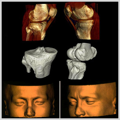
3D CT Reconstruction
Three-dimensional imaging, performed by Advanced Imaging Center's Picker Dual Slice Helical CT that acquires data volumetrically, allows computer processing to display various organs and disease processes three dimensionally. This is now feasible because the volumetric scan data is free of motion artifact and the low cost of computer power combined with three-dimensional software can create images previously unobtainable. This process has many applications. Click here for more details.
Click Here to Read more
4D-Multiplanar CT
Three-dimensional imaging, performed by Advanced Imaging Center's Picker Dual Slice Helical CT that acquires data volumetrically, allows computer processing to display various organs and disease processes three dimensionally. This is now feasible because the volumetric scan data is free of motion artifact and the low cost of computer power combined with three-dimensional software can create images previously unobtainable. This process has many applications.
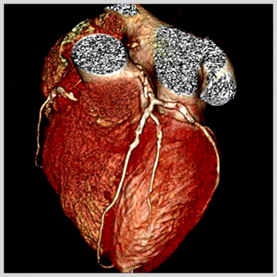
CT Angiography
The imaging of blood vessels in detail comparable to catheter angiography at a fraction of the time and cost is a remarkable advance. This technique will replace, to a great extent, invasive angiography.
Musculoskeletal
Imaging of bones and joints three dimensionally allows a different perspective for understanding pathology such as complex fractures. It is very helpful for surgical planning.
Therapy Planning
Volume rendering is the most recent development. It is a method by which the viewer is provided with a three-dimensional perspective of hollow organs. This technique known as virtual endoscopy will be useful to image the trachea, bronchi, and the colon. Virtual endoscopy of the colon is performed after a thorough colon cleansing and distention of the colon with air. The helical CT scan data is then processed to allow the viewer a three dimensional view of the inside of the colon similar to that from an endoscope. This may someday play a role in colon cancer screening.
Volume Rendering
Volume rendering is the most recent development. It is a method by which the viewer is provided with a three-dimensional perspective of hollow organs. This technique known as virtual endoscopy will be useful to image the trachea, bronchi, and the colon. Virtual endoscopy of the colon is performed after a thorough colon cleansing and distention of the colon with air. The helical CT scan data is then processed to allow the viewer a three dimensional view of the inside of the colon similar to that from an endoscope. This may someday play a role in colon cancer screening.
All Inclusive" Comprehensive Imaging
Three-dimensional helical CT in many cases can now provide all the information necessary for definitive treatment, which previously required two or more radiological studies. Its ability to display both the pathology within an organ and blood supply to the organ is unique and provides the most information for the least cost.


Health Scan
Full Body Health Scan (FBHS) is an examination of the body by utilization of an ultra-fast helical CT scanner for cancer screening and coronary calcification scoring. This WHOLE BODY SCAN screens for tumors in the body and detects the amount of calcium build up in heart arteries. The purpose of the scanning is to detect lung cancer, abdominal or pelvic cancer, and coronary artery disease. The results of the scan are achieved quickly and painlessly. Click here for more details.
Click Here to Read more
WHAT IS FULL BODY HEALTH SCAN?
Full Body Health Scan (FBHS) is an examination of the body by utilization of an ultra-fast helical CT scanner for cancer screening and coronary calcification scoring. This WHOLE BODY SCAN screens for tumors in the body and detects the amount of calcium build up in heart arteries.
The purpose of the scanning is to detect lung cancer, abdominal or pelvic cancer, and coronary artery disease. The results of the scan are achieved quickly and painlessly. An analysis of the scan outcome enables the radiologist to guide you and your physician regarding lifestyle and/or diet modifications or treatment to optimize your health.
AIC's coronary calcium scoring measures the amount of calcium in the coronary arteries… a measure of coronary artery disease. This program can tell you if you are at risk for a heart attack. For anyone over the age of 40 and those who smoke, have a family history of heart problems or are currently diagnosed with high blood pressure, diabetes or other heart related diseases, scan-health is something you should look in to.
WHY THIS SCAN IS IMPORTANT
Your future depends on the present. This non-invasive tool can give you the tools to reveal the true status of your health. One office visit. One scan. One consultation. In one single and painless visit is all it takes. No need to schedule multiple appointments, or waiting endless days to hear about the results of the tests done. Now YOU can take destiny in your hands and potentially save your life.
WHY THIS SCAN IS IMPORTANT
- - Arrive at Advanced Imaging Center approximately 15 minutes before your scheduled appointment. Park in the parking lot.
- - Complete the medical questionnaire (5-10 minutes).
- - You will lie on the table and in several short breath-holds, the scanner will take the images.
- - It will take 5-10 minutes to complete the scan.
- - The unique innovative technology of the ultra-fast, helical CT scanner delivers high-resolution images in a fraction of the time of conventional CT scanners.
- - Radiation is less than that of a conventional CT scanner.
- - Painless, non -invasive and non-claustrophobic.
WHAT TO EXPECT AFTER YOUR VISIT
- - Schedule an in-person consultation with the radiologist (15-20 minutes.)
- - An opportunity to view the scan, ask questions, discuss abnormalities.
- - A detailed written report will follow with a copy sent to your physician within 2 -3 days.
SCREENING CORONARY SCAN
- - Screening coronary artery calcium scoring.
- - 1.5 million Americans died from Cardiovascular diseases and Cancer last year.
- - Some of these deaths could have been prevented!
SCREENING TUMOR SCAN
Cancer is a group of diseases characterized by uncontrollable growth and spread of abnormal cells. If the spread is not controlled, it can result in death. Many cancers are cured if detected and treated promptly. Many can be prevented by lifestyle changes.
What question can Full Body Health Scan answer?
- - Is a tumor present?
- - Has the cancer spread?
- - Is my cancer treatment effective?
- - Has the cancer recurred?
What are the risk factors?
- - Family history of cancer
- - Tobacco and alcohol use
- - Environmental exposure nutrition and diet
- - Age, gender, ethnicity.
According to the American Cancer Society
"Cancer kills more people annually than AIDS, accidents and homicide combined. Many of the deaths could have been prevented through increased cancer prevention and screening and improved access to medical care."
Full Body Health Scan can mean the difference between living life to the fullest or becoming a victim of a disease
The Full Body Health Scan is a non-invasive CT scan of the body performed on our Dual Slice Helical CT. A specially trained radiologist examines your cardiovascular system, lungs, and major organs of the abdomen and pelvis to detect the earliest stages of disease, including coronary artery disease and lung cancer.
Results from this rapid and painless scan indicate whether or not hidden disease exists and enables the radiologist to guide you and your physician on possible lifestyle/diet modifications, or treatment, to optimize your health.
Your future depends on the present. The most expensive cost in medicine has proven to be the unknown. Advanced Imaging Center's Full Body Health Scan, one of the most powerful, non-invasive tools to reveal the unknown, is available to your and your family. Now YOU can take destiny in hand to potentially save your own life.
In one single and painless visit, multiple diagnostic tests are run simultaneously eliminating the need for you to return to a facility repeatedly to learn the status of your health. One office visit. One scan. One in-person consultation with the same physician who interprets the scan to give you an opportunity to view the scan, ask questions, and discuss abnormalities. A comprehensive, written report will follow, with a copy to your physician within 2-3 days.
If you would like to receive more information about the Full Body Health Scan, or if you would like one of our staff members to contact you, please call us for information or to set up an appointment.
The Full Body Health Scan is very accurate in detecting the following diseases:
- Chest
- .
- - Coronary artery disease
- - Lung cancer, emphysema
- - Thoracic aorta calcification, dilatation
- - Pericardial effusion
- - Heart valve calcification
- Abdomen
- .
- - "Silent tumors" of the liver, pancreas, kidneys, adrenal glands, and lymph nodes
- - Stomach tumors and inflammation
- - Liver cysts, infection, cirrhosis
- - Abdominal aortic aneurysm
- - Gallstones
- - Kidney stones
- Pelvis
- .
- - Male
- - Prostatic enlargement
- - Bladder tumors
- - Female
- - Ovarian and uterine tumors
- - Bladder tumors
- Skeletal System
- .
- - Bone demineralization (osteoporosis)
- - Bone tumors
- - Spine and hip arthritis and Degenerative disease
- - WHAT IS CORONARY ARTERY DISEASE (CAD)?
CAD is narrowing or blockage of the coronary arteries secondary to atherosclerotic plaque ("hardening of the arteries"). Most plaque calcifies, and the greater the amount of calcium (which we can measure) the more severe the plaque and blockage. The coronary arteries supply blood to the heart muscle (myocardium). If the coronary artery is obstructed, blood flow to the heart muscle is decreased or stopped, resulting in a heart attack (myocardial infarct).
What Do the Results of the Full Body Health Scan Tell Me About my Cardiovascular System?
A score is computed based on the amount of calcification detected. Your score is an accurate predictor of the degree of narrowing of coronary arteries and the likelihood of a future coronary event (heart attack). If no calcified plaque is detected, the risk of CAD can be virtually excluded.
What Are the Risk Factors According to the American Heart Association?
- - Non-modifiable Risk Factors
- - Men older than 45
- - Postmenopausal women
- - Family history of heart disease
- - Modifiable Risk Factors
- - Smoking
- - Obesity
- - Sedentary lifestyle
- - Hypertension
- - Diabetes
- - Elevated LDL, low HDL
According to the American Heart Association:
- - CAD is the number one killer of American men and women.
- - Every 33 seconds someone in the United States dies from heart disease. 30% have no apparent risk factors.
- - American Heart Association, Heart and Stroke Statistical Update, 1998.
WHAT IS CANCER?
Cancer is a group of diseases characterized by uncontrolled growth and spread of abnormal cells. If the spread is not controlled, it can result in death. However, many cancers can be cured if detected and treated promptly, and many others can be prevented by lifestyle changes, especially avoidance of tobacco.
What Questions Can the Full Body Health Scan Answer with a High Degree of Accuracy?
- - Is a silent (asymptomatic) tumor present?
- - Has the cancer spread?
- - Is my cancer treatment effective?
- - Has the cancer recurred?
What are the Risk Factors According to the American Cancer Society?
- - Non-modifiable Risk Factors
- - Family history of cancer
- - Age
- - Gender
- - Ethnicity
- - Modifiable Risk Factors
- - Tobacco
- - Alcohol abuse
- - Environmental exposure
- - Nutrition and diet
- - According to the American Cancer Society
According to the American Cancer Society
"Cancer affects Americans of all racial and ethnic groups and kills more people annually than AIDS, accidents, and homicides combined. In 1997 alone, the American Cancer Society estimates that 560,000 cancer deaths will occur and 1,382,400 new cases will be diagnosed. Many of these deaths could be prevented through increased cancer prevention and screening, and improved access to medical care."
American Cancer Society's Annual Report, Cancer Facts and Figures, 1997.


Coronary Calicium Scoring
Coronary artery disease (CAD) is the leading cause of death in the United States. Each year 30-50% of the 1.5 million American men and women who have a heart attack (myocardial infarct) die as a result. Most of these occur in people who've had no previous symptoms or warning. Men are at greater risk for heart attack at certain times of their lives, but overall men and women die at equal rates from CAD. Click here for more details.
Click Here to Read more
Q. What is Coronary Calcification Scoring?
A. Coronary calcification scoring or cardiac scoring is a CT technique to determine the amount of calcium build up in the coronary arteries. Coronary artery calcification is a specific marker for coronary atherosclerosis. The amount of calcification correlates with severity of coronary atherosclerosis and the probability of obstructive disease.
Q. How is it performed?
A. The scan is performed on an ultrafast CT (either helical or electron beam CT with similar accuracy) in one breathhold. At AIC a 16-slice ultrafast multi-slice, multi-detector helical CT is used, and the whole procedure takes just a few minutes.
Q. What happens after the scan?
A. The data are processed via a special cardiac scoring software package. A radiologist then evaluates the images and puts region of interests (ROI's) on the calcified coronary arteries. At the end, individual scores for four arteries (left main, LAD, circumflex, and right coronary) and a total score are calculated. The total score falls under one of the following categories:
- 0-1: NO CALCIFICATION (extremely low likelihood for obstructive coronary disease);
- 1-10: MINIMAL CALCIFICATION;
- 11-100: SMALL AMOUNT CALCIFICATION;
- 101-400: MODERATE CALCIFICATION;
- 400: LARGE AMOUNT CALCIFICATION (high likelihood for extensive coronary atherosclerosis).
Q. What's the accuracy of the test?
A. It has a nearly 100% sensitivity for calcifications and nearly 100% negative predictive value for future coronary events. The positive predictive value ranges from 50 to 80. A zero or very low score implies virtually no coronary obstructive disease with the exception occurring in young patients who smoke (soft plaques). A high score indicates a significant plaque burden and risk for future cardiovascular event. It should be understood that calcification is not site specific for stenosis but rather indicates the extent of atherosclerosis in the coronary arteries overall. The score may be used as benchmark to measure subsequent disease development or assess preventive programs.
Q. Who should get this test?
A. Individuals who have any of the following: history of smoking, diabetes, hypertension, hypercholesterolemia, family history of coronary artery disease, obesity, sedentary lifestyle, high level of stress, atypical chest pain, asymptomatic males over 45 and females over 55 years of age.
Coronary artery disease (CAD)
Coronary artery disease (CAD) is the leading cause of death in the United States. Each year 30-50% of the 1.5 million American men and women who have a heart attack (myocardial infarct) die as a result. Most of these occur in people who've had no previous symptoms or warning. Men are at greater risk for heart attack at certain times of their lives, but overall men and women die at equal rates from CAD.
Coronary artery calcification scoring is a non-invasive CT scan of the heart performed on our 16-slice Helical CT. The scan detects and quantifies calcified atherosclerotic plaque in the coronary arteries. A score is computed based on the amount of calcification detected. Your score is an accurate predictor of the degree of narrowing of the coronary arteries and the likelihood of a future coronary event (heart attack). The radiologist, an imaging specialist, will interpret the scans and send a report to you and your physician.
The American Heart Association has identified the following risk factors:
- - Men over age 45
- - Women over age 55
- - Elevated LDL cholesterol
- - Low HDL cholesterol
- - Family history of coronary artery disease
- - Smoking
- - Obesity
- - Sedentary life style
- - High blood pressure
- - Diabetes
If you are a male over 45 or a female over 55 and have one or more of these risk factors.
CORONARY CALCIFICATION SCORING WILL BE USEFUL TO YOU!
500,000 Americans die from coronary artery disease (heart attack) yearly. Most have no warning prior to their death! Early detection of calcified atherosclerotic plaque can prompt preventive action to minimize risk of heart attack or direct you to seek medical evaluation for further testing. Coronary atherosclerosis can be slowed, stopped, and possibly reversed before artery blockage results in heart muscle damage or death.
Remember, in cardiac disease, PREVENTION could mean everything!
Coronary Artery Calcification Scoring has clinical value:
- - To determine if patients with chronic atypical chest pain have coronary atherosclerosis.
- - To screen asymptomatic patients in order to stratify their risk of coronary disease and future cardiac events.
- - To exclude the presence of coronary artery disease in women over 60 years of age.
- - To determine if patients with equivocal stress or thallium test have coronary artery plaque before proceeding to coronary angiography.
- - To identify postmenopausal women with low risk for coronary disease who are therefore less likely to benefit from cardioprotective effects of hormone replacement therapy.
- - To determine if a dilated cardiomyopathy is secondary to coronary artery disease or not.
- - To simplify the pre-operative cardiac clearance of women over age 60.
- - To follow the progression of coronary artery plaque non-invasively.
- - Read selected journal references for Coronary Artery Calcification Scoring.
Q & A regarding AIC's Ultrafast, multidetector Helical CT
Q. Can you tell me about the new helical CT at AIC?
A. Certainly. It is a 16-slice CT. The new helical CT has replaced our old dual-slice CT. It is a multi-slice (multi-detector) CT capable of simultaneously scanning 16 slices evry 0.4 second (or 40 slices per second), thus increasing the speed of CT scanning by at least a factor of 40 or more. It is the fastest CT in the Antelope Valley area.
Q. What does ultrafast CT allow you to do?
A. Here's a summary (* denotes AIC exclusive):
- 1- Fast high-resolution (1-2 mm) routine imaging of the neck, chest, abdomen, and pelvis.
- 2- *Fast high-res (0.5 mm) imaging of the IAC's/Temporal bones.
- 3- *Fast ultra-high-resolution (0.5 mm) imaging of the bones for 4D isotropic reconstruction.
- 4- 3D and 4D CT Angiography (CTA): aorta, pulmonary arteries, runoffs, brain.
- 5- Coronary artery calcification scoring (excellent non-invasive screening test).
- 6- Virtual endoscopy (colonoscopy, bronchoscopy, and endovascular angioscopy with CT!)
Q. What does coronary calcification scoring tell you?
A. Coronary scoring is a safe, noninvasive and fast screening CT technique that scans the heart in a few seconds and gives a score based on the amount of calcium build up in the coronary arteries. The score is a good predictor of future coronary events. For instance, it has a 100% negative predictive value!
Q. Can you tell me more about the new workstation at AIC?
A. Certainly. The new workstation is a state-of-the-art silicon graphics workstation linked to all our modalities including Open MRI, high-field MRI, Helical CT, and Nuclear med (SPECT/PET). It is a powerful and sophisticated computer capable of amazing multiplanar and 4D reconstruction, fusion of images from different modalities, 4D virtual endoscopy, dental scan, 3D/4D CT Angiography (CTA), just to name a few. It is simply technology at its best.
Q. What is virtual endoscopy?
A. This noninvasive technique is one of the hottest areas in CT today that allows 4D visualization of hollow organs (colon, bronchus, etc.) similar to video endoscopy. This is only available at AIC.
Q. I have not heard of image Fusion. Can you explain?
A. Of course. Image fusion is a sophisticated software utilized by our workstation that allows fusion of images from different modalities (e.g., CT, MRI, SPECT, PET) or fusion of images from the same modality at different times (to evaluate for growth). For example, a physiologic/metabolic SPECT or PET image can be combined with an anatomic CT or MRI image to provide an anatomico-physiologic image.


High Field MRI
It is capable of performing the most complex MRI studies, including tasks not available anywhere else in the area (e.g., Perfusion/Diffusion study of the brain for early stroke detection; Breast MRI for implants and cancer; CSF Flow studies; Contrast-enhanced peripheral MRA with power injector; MRCP [MR version of ERCP]; MR Myelography; ultra fast single-breath hold abdominal scanning; MR neurogram; high-performance gradients, thus ultra fast (sub second) and ultra high-res (1024 matrix); just to name a few). Click here for more details.
Click Here to Read more
Advanced Imaging Center also provides the first short-bore, wide flared high-field (1.5 Tesla) MRI in the valley, the first one of its kind in Southern California, which is the most advanced MRI system in the world (top right). Same field strength as the closed MRI's (i.e., 1.5 Tesla) but with open features, this Siemens Symphony MRI with high-performance gradients is unmatched in the area.
It is capable of performing the most complex MRI studies, including tasks not available anywhere else in the area (e.g., Perfusion/Diffusion study of the brain for early stroke detection; Breast MRI for implants and cancer; CSF Flow studies; Contrast-enhanced peripheral MRA with power injector; MRCP [MR version of ERCP]; MR Myelography; ultra fast single-breath hold abdominal scanning; MR neurogram; high-performance gradients, thus ultra fast (sub second) and ultra high-res (1024 matrix); just to name a few). AIC is the only facility in the Antelope Valley that is MRI certified (accredited by the American College of Radiology).
MRI, magnetic resonance imaging, is a method used by physicians to visualize internal organs of the human body and obtain diagnostic information. These images are produced without the use of radiation. MRI is a noninvasive procedure and there are no known side or after effects. The procedure is painless. You won't see or feel anything during the exam. A faint knocking sound will be heard which is the imaging process in operation.
MRI utilizes the physical properties of magnetic fields, radio waves, and computers to generate images of the body in any plane. The technique is an established, accurate diagnostic tool. It can quickly and noninvasively provide accurate diagnosis for your physician, which in some situations will reduce the need for exploratory surgery or other diagnostic procedures, which may have greater risk.
The benefits of magnetic resonance imaging are vast and new applications are continually being developed through ongoing research. MRI imaging is used for virtually all parts of the body. It is the primary imaging modality for evaluation of diseases of the brain and spine. It is effective in depicting abnormalities of the eye, paranasal sinuses, throat, salivary glands, and the thyroid.
MRI is the method of choice for imaging of the musculoskeletal system and is widely used for evaluation of the shoulder, elbow, wrist, hip, knee, and ankle. It can also accurately depict abnormalities within the bone marrow. It has many applications in the cardiovascular system. The display of blood vessels known as MR angiography is an accurate, noninvasive means of obtaining information about arteries of the head, neck, and body.
It is also effective in demonstrating congenital cardiac abnormalities. MRI is also important in evaluation of the organs of the abdomen and pelvis including the liver, pancreas, kidneys, ovaries, uterus, and prostate.


3D Virtual Colonoscopy (VC)
Virtual Colonoscopy (VC) is a new method that allows 3-D visualization of the large bowel (colon) to detect polyps and cancers. Polyps are small growths in the colon that may become cancerous if they are not removed. Virtual Colonoscopy is a recently developed technique that uses a fast multi-slice helical CT scanner (MSCT) and computer 3D virtual reality software to look inside the bowel lumen without having to insert a long scope (Conventional Colonoscopy) or having to fill the colon with liquid barium (Barium Enema). Click here for more details.
Click Here to Read more
Virtual Colonoscopy (VC) is a new method that allows 3-D visualization of the large bowel (colon) to detect polyps and cancers. Polyps are small growths in the colon that may become cancerous if they are not removed. Virtual Colonoscopy is a recently developed technique that uses a fast multi-slice helical CT scanner (MSCT) and computer 3D virtual reality software to look inside the bowel lumen without having to insert a long scope (Conventional Colonoscopy) or having to fill the colon with liquid barium (Barium Enema).
Patients need a cleansing preparation of their bowel prior to the test. The actual VC procedure will begin by having a small flexible rubber tube placed in the rectum, so that air can be introduced. A helical CT scan is then performed while the patient lies comfortably on the back (supine) and then on the stomach (prone). The total time required for the study is around 15 minutes. Because sedation is not required, patients are free to leave the CT suite immediately without the need for observation or recovery. Patients can resume normal activities immediately after the procedure and can eat, work or drive without a delay. When air is introduced in the colon, some patients experience minimal temporary abdominal cramping or "gas pains."
ACCURACY
Research has shown that Virtual Colonoscopy is better able to see polyps than Barium Enema and is nearly as accurate as Conventional Colonoscopy. Fig. 1 shows a polyp and Fig. 2 a flat cancer seen on 3 techniques: 2D axial CT (Fig. 1a, 2a-b), 3D VC (Fig. 1b, 2c), and conventional colonoscopy (Fig. 1c, 2d). Most patients report that the Virtual Colonoscopy technique is more comfortable than either Barium Enema or Conventional Colonoscopy. Studies suggest a very high sensitivity and specificity (96%) for the detection of polyps 1 cm or greater. Such polyps have significant malignant potential. Sensitivity for polyps less than 1 cm is significantly less. Although controversy exists as to the definition of a "significant" polyp with regard to size, polyps smaller than a 1 cm in size have a low probability of malignancy and the likelihood of any single lesion progressing to cancer is also small.
INDICATIONS
Indications for VC include screening for polyps, incomplete or failed colonoscopy, and preoperative assessment of the colon proximal to an occlusive cancer (defined as a tumor that cannot be traversed endoscopically). Virtual colonoscopy excels in the evaluation of the ascending colon, particularly the cecum due to the degree of distension achievable and the typical lack of spasm or muscular hypertrophy, which is seen in the sigmoid colon. It is therefore a useful complement to an incomplete colonoscopy. Any questionable abnormality will warrant a conventional colonoscopy procedure, thus resulting in GI referrals. VC may become a screening tool for detecting colorectal neoplasia, potentially supplanting Conventional Colonoscopy as a tool for detecting lesions. Increased screening, with increased detection, should decrease the incidence of colorectal cancer, as premalignant growths can be found at an earlier stage. About 5% of colon cancers (flat cancers) arise from normal mucosa and not from polyps and are difficult to identify on conventional and virtual colonoscopy, while axial helical CT images improve detection of such cancers.
HEALTHSCAN PROGRAM
AIC has been offering for some time a screening total-body scan program called HealthScan, which includes a screening helical CT of the torso, helical CT coronary calcium scoring, and more optional 3D virtual colonoscopy.


New 64 Slice CT
Advanced Imaging Center's Open MRI features the Hitachi AIRIS Open MRI, the most advanced open MRI system. It was the first Open MRI in the Antelope Valley and still the only high-quality one. With our Open MRI you can enjoy the benefits of MRI without the fear of confinement.
This state-of-the-art open MRI has a unique, "open on all 4 sides" design which is non-claustrophobic. Click here for more details.
Click Here to Read more


16-Slice CT
It is with great pleasure and excitement to announce that AIC will be installing a brand new multi-slice CT scanner in May 2003. As you may recall, AIC was the first to introduce Multi-Slice CT (MSCT) to the valley in 1999 with its dual-slice CT, then state-of-the-art. Now we take pride in introducing the next generation in CT technology to the valley. This new 16-slice MSCT scanner is the most advanced CT scanner in the world and the first and oly one of it's kind in the valley. Click here for more details.
Click Here to Read more
It is also effective in demonstrating congenital cardiac abnormalities. MRI is also important in evaluation of the organs of the abdomen and pelvis including the liver, pancreas, kidneys, ovaries, uterus, and prostate.


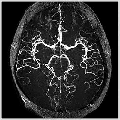
MR Angiography
MR Angiography is a noninvasive method to evaluate the patency of blood vessels without resorting to invasive conventional x-ray angiography. The latter requires insertion of a catheter into an artery (usually in the groin) and carries a certain amount of risk (infection, vessel rupture, hematoma, occlusion, embolism, allergies to iodine dye, etc.) Unlike x-ray angiogram, MR angiogram (MRA) is performed either with no dye or with intravenous (as opposed to intra-arterial) injection in an arm vein. The contrast or dye used in MRA has no iodine in it and is much safer than x-ray dye. MRA can be performed in the head, neck (carotids), body, arm and legs.
Click Here to Read more
MR Angiography is a noninvasive method to evaluate the patency of blood vessels without resorting to invasive conventional x-ray angiography. The latter requires insertion of a catheter into an artery (usually in the groin) and carries a certain amount of risk (infection, vessel rupture, hematoma, occlusion, embolism, allergies to iodine dye, etc.)
Unlike x-ray angiogram, MR angiogram (MRA) is performed either with no dye or with intravenous (as opposed to intra-arterial) injection in an arm vein. The contrast or dye used in MRA has no iodine in it and is much safer than x-ray dye.
MRA can be performed in the head, neck (carotids), body, arm and legs.
At Advanced Imaging Center, MRA can be performed on both the OPEN MRI and the high-field MRI. The latter has more MRA applications, including contrast-enhanced MRA of the carotids, aorta, pulmonary arteries, renal arteries, and extremities, not possible on an open system as they required very high resolution and speed.
The high-field Siemens Symphony MRI system at AIC has the most sophisticated MRA package available anywhere such as:
- - 20-second Gadolinium-enhanced MRA of the carotids utilizing an MR-compatible power injector;
- - 30-second Gadolinium-enhanced body MRA (pulmonary arteries, renal arteries, aorta, mesenteric angio;
- - 30-second Gadolinium-enhanced peripheral MRA (run-offs utilizing a new automatic table-moving feature and a power injector with accuracy similar to or better than X-ray angio).
- - Exquisite ultra-resolution MRA of the circle of Willis and the rest of the brain using Advanced MRA.
Contrast-Enhanced Subtraction MR Angiography (CE-MRA): State-of-the-art
CE-MRA of the aortic arch and great vessels (top left) into the brain, renal arteries (top), selective carotid bifurcation (left) showing ICA/ECA stenosis, and 3-section run-off (right) showing multiple stenoses, including long-segment bilateral SFA stenoses.
Q. What is Contrast-Enhanced MR Angiography (CE-MRA)?
A: CE-MRA is a fairly new, non-invasive MR technique that utilizes ultra fast MR sequences to obtain exquisite MRA images. These images are obtained during intravenous administration of only 20-30 cc of Gadolinium using an MR-compatible power injector. A bolus-tracking software tracks the bolus of contrast for optimal timing. Subtraction techniques may also be utilized for improved tissue contrast. Thus, a high-end scanner is required to perform the procedure.
Q. What are some applications?
A: Carotids, brain, aorta, renals, pulmonary, run off, etc.
Q. How long does it take to do the carotids, for example?
A: The actual scan is performed in one breath hold (about 20 seconds), but of course it takes some time to prepare the patient and set up the protocols. The whole procedure can be performed in ½ hour. 4D reconstruction is then performed.
Q. What is the accuracy of the test?
A: It is extremely accurate when performed properly, and it correlates well with x-ray angiography and can replace it in many instances. Some patients may undergo angioplasty or surgery based on CE-MRA without the need for conventional angiography. 4D reconstruction allows visualization at multiple angles. Best of all, it is non-invasive!


CT Angiography
Three dimensional imaging, performed by our Toshiba 16-slice Helical CT which acquires data volumetrically, allows computer processing to display various organs and disease processes three dimensionally. This is now feasible because the volumetric scan data is free of motion artifact and the low cost of computer power combined with three dimensional software can create images previously unobtainable. This process has many applications. Click here for more details.
Click Here to Read more
Three dimensional imaging, performed by our Toshiba 16-slice Helical CT which acquires data volumetrically, allows computer processing to display various organs and disease processes three dimensionally. This is now feasible because the volumetric scan data is free of motion artifact and the low cost of computer power combined with three dimensional software can create images previously unobtainable. This process has many applications.
CT Angiography
The imaging of blood vessels in detail comparable to catheter angiography at a fraction of the time and cost is a remarkable advance. This technique will replace, to a great extent, invasive angiography.
Musculoskeletal
Imaging of bones and joints three dimensionally allows a different perspective for understanding pathology such as complex fractures. It is very helpful for surgical planning.
Therapy Planning
Surgical and radiation treatment procedures are complex three dimensional problems. Three dimensional rendering of CT data is helpful for surgical planning and for the radiation oncologist. For both, understanding the relationship of tumors to normal tissue and blood vessels is critical. These imaging techniques are providing information not previously obtainable.
Volume Rendering
Volume rendering is the most recent development. It is a method by which the viewer is provided with a three dimensional perspective of hollow organs. This technique known as virtual endoscopy will be useful to image the trachea, bronchi, and the colon. Virtual endoscopy of the colon is performed after a thorough colon cleansing and distention of the colon with air. The helical CT scan data is then processed to allow the viewer a three dimensional view of the inside of the colon similar to that from an endoscope. This may someday play a role in colon cancer screening.
All Inclusive" Comprehensive Imaging
Three dimensional helical CT in many cases can now provide all the information necessary for definitive treatment which previously required two or more radiological studies. Its ability to display both the pathology within an organ and blood supply to the organ is unique and provides the most information for the least cost.
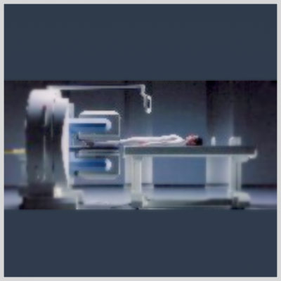
Nuclear Medecine/Spect
Q. Can you tell me more about the new NUCLEAR/SPECT/PET camera at AIC?
A. The Nuclear scanner at AIC (shown above) is the most sophisticated in the Antelope Valley area. It is a top-of-the-line Siemens state-of-the-art dual-head gamma camera that can perform full range of Nuclear Medicine scans including SPECT scans as well as PET scans. Click here for more details.
Click Here to Read more
AIC has the valley's most sophisticated nuclear camera.
Q. Can you tell me more about the new NUCLEAR/SPECT/PET camera at AIC?
A. The Nuclear scanner at AIC (shown above) is the most sophisticated in the Antelope Valley area. It is a top-of-the-line Siemens state-of-the-art dual-head gamma camera that can perform full range of Nuclear Medicine scans including SPECT scans as well as PET scans.
Q. What types of Nuclear studies are offered at AIC?
A. Full nuclear services are offered including bone scans, HIDA scans, V/Q scans, thyroid scans, Indium white cell scans, Gallium scans, brain SPECT, scintilymphangiography, cardiac scans (thallium and Sestamibi SPECT, etc.).
Q. What is SPECT and what are some of the indications?
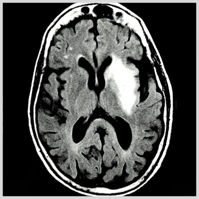
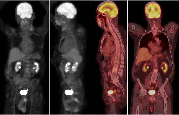
A. SPECT stands for Single Photon Emission Computerized Tomography. SPECT is to planar nuclear scanning as CT (CAT scan) is to planar x-rays. SPECT images are tomographic slices through the region of interest. It is more sensitive than planar scanning due to its finer thickness. Multiplanar imaging (coronal, sagittal, axial) is available. SPECT is now the standard technique in cardiac imaging, but has broad applications such as in bone scanning. Images on the right show a coronal CT reformation of the spine demonstrating a sclerotic lesion in T12 vertebral body and the corresponding coronal SPECT image clearly showing increased activity in T12 (arrows). The planar whole body bone scan (not shown) was negative failing to show the T12 lesion.
Q. What is a PET scan and what are some of the indications?
A. PET stands for Positron Emission Tomography. PET imaging uses a glucose analogue agent called F18-Fluoro-Deoxy-Glucose or FDG. In the body, PET is used in oncology to detect malignancy since malignant cells demonstrate higher glucose metabolism. Thus it can differentiate between malignant and benign tumors and between recurrent tumor and scar tissue/radiation changes, etc. In the brain, it can be used for malignancy as well as Seizures/Epilepsy, Alzheimer and other Dementias, Schizophrenia, Depression, Attention Deficit Disorder (ADD), etc. The images on the right show a reformatted coronal CT image with the corresponding coronal PET image of an unsuspected destructive metastasis to the left upper rib cage (arrows).


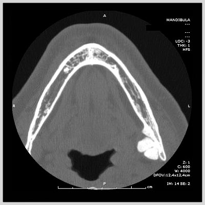
Dental Scan
The study is performed on our multi-slice helical CT scanner. One-millimeter scans are performed through the mandible and/or maxilla. The scans are then sent to an advanced workstation where they are reconstructed to display both panoramic and transverse views. The scan images are then printed in true anatomical scale on full size radiographic film. The whole process is very quick. Films are ready for pickup or delivery that same day, often within thirty minutes. Click here for more details.
Click Here to Read more
Advanced Imaging Center is proud to introduce CT scanning for dental implant planning. Using a scan protocol reviewed at Yale University, we provide you with vital information about your patient’s dental anatomy.
The study is performed on our multislice helical CT scanner. One-millimeter scans are performed through the mandible and/or maxilla. The scans are then sent to an advanced workstation where they are reconstructed to display both panoramic and transverse views. The scan images are then printed in true anatomical scale on full size radiographic film. The whole process is very quick. Films are ready for pickup or delivery that same day, often within thirty minutes.
Some Highlights:
- - Same day scheduling and turn around
- - Convenient delivery of films to your office
- - True anatomic scale images
- - Actual radiographic film, no photo or inkjet paper images
- - Also available, temporomandibular joint (TMJ) MRI


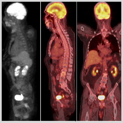
PET-CT
Presenting our new Simens Biograph 40 slice PET-CT, which is yet another first to the Antelope Valley, and is the only one of it's kind within a 100 mile radius. Our 40 slice PET-CT is capable of performing a Coronary CTA. The Biograph is also capable of performing two scans at once time which decreases the scan time.
Advanced Imaging Center has the only PET-CT in the area. Click here for details.
Click Here to Read more
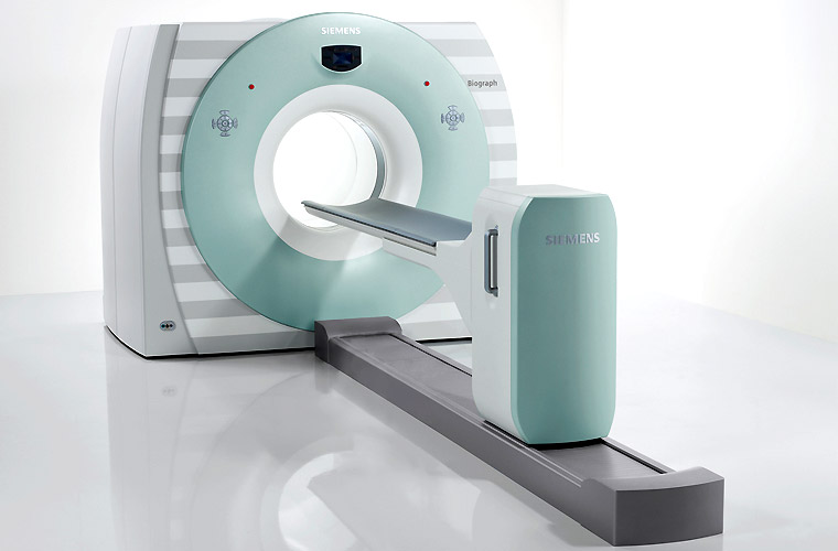
Presenting our new Simens Biograph 40 slice PET-CT, which is yet another first to the Antelope Valley, and is the only one of it's kind within a 100 mile radius. Our 40 slice PET-CT is capable of performing a Coronary CTA. The Biograph is also capable of performing two scans at once time which decreases the scan time.
Patients will appreciate the faster scan times, and doctors will be amazed at the high quality of the 2D and 3D images it produces.
Advanced Imaging Center has the only PET-CT in the area.


Dexa Bone Densitometry
Up to 30% of elderly people with hip fractures die within 6 months of their injury. The difference in sex distribution in osteoporosis is especially significant as women who are 65 years of age or older represent the fastest growing segment of the population in the United States. The worldwide increase in life expectancy will most likely result in an accompanying rise in the prevalence of osteoporotic fractures of all kinds over the next decades. Click here for more details.
Click Here to Read more
Advanced Imaging Center offers one of the world's most sophisticated DEXA scanners.
DEXA (stands for Dual Energy X-ray Absorptiometry) is the safest and most accurate method to measure Bone Mineral Density, an overall measure of how much calcium there is in the bones.
A low level of BMD below a certain threshold (below 2 standard deviations from peak bone mass) indicates osteoporosis.
DEXA provides low cost, state-of-the-art Bone Densitometry with insignificant radiation, unlike CT or nuclear bone densitometry.
Osteoporosis is a common disorder affecting a large number of adults. In the United States more than 25 million people are afflicted, particularly women. Many suffer disabling fractures of the spine, which is the most common site of involvement. Osteoporosis is believed to be responsible for about 1.3 million fractures annually including more than 500,000 spine, 250,000 hip, and 240,000 wrist fractures.
Up to 30% of elderly people with hip fractures die within 6 months of their injury. The difference in sex distribution in osteoporosis is especially significant as women who are 65 years of age or older represent the fastest growing segment of the population in the United States. The worldwide increase in life expectancy will most likely result in an accompanying rise in the prevalence of osteoporotic fractures of all kinds over the next decades.
During the past decades, osteoporosis, called the "silent epidemic," has gained increased attention. The involvement chiefly of women and the insidious loss of bone manifested primarily as "crush fractures" of the spine, hip and wrist are widely known facts. Public awareness of this disorder also has been heightened by the resulting increase in health care expenditure that is currently estimated to be in excess of 7 billion dollars.
Indications for DEXA:
- - Patients receiving long term glucocorticoid therapy.
- - Patients with primary asymptomatic hyperparathyroidism
- - Patients at high risk for osteoporosis such as amenorrhea, anorexia nervosa or alcoholism
- - Patients with atraumatic fractures, disuse atrophy, and similar conditions
- - Assessment of early postmenopausal bone loss as an indication to initiate estrogen replacement therapy
- - Diagnosis of osteoporosis suspected from radiographic findings or from clinical risk factors
- - Serial assessment of bone density, i.e., during treatment for osteoporosis or in anticipation of rapid bone loss
A variety of metabolic disorders such as hyperparathyroidism, renal insufficiency, Cushing's syndrome, and amenorrhea in premenopausal women as well as chronic immobilization and chronic steroid or thyroid therapy are known to influence calcium metabolism and may affect the skeleton adversely. In these cases of secondary osteoporosis, bone density measurements are of particular importance because they may prompt therapeutic decisions such as reduction in medication or surgery.
Bone turnover increases significantly at menopause with a greater increase in bone resorption than bone formation resulting in accelerated loss of bone. One-third to one-half of bone loss in women may be attributable to the loss of ovarian function. Several studies have established the bone mass-preserving effect of estrogen therapy; if begun soon after menopause it reduces the subsequent rate of vertebral fractures by 50%. The benefits derived from estrogen therapy clearly seem to outweigh its adverse effects. However, it is unacceptable for many women and the level of bone mineral density at menopause and the magnitude of subsequent loss are important considerations in assessing the future risk of fracture and a decision to begin prophylaxis can be based on such considerations.
Serial measurement of bone density is accurate, providing guidance for clinical treatment. Measurements every 1-2 years are useful depending on the disease process. Studies have shown large annual loss of bone density from sites rich in trabecular bone in patients receiving high dose steroids. Similarly, large annual gains of bone have been observed in osteoporotic patients receiving treatment with a variety of therapeutic agents such as calcitonin, fluoride, biphosphonates, or parathyroid hormone. Considering the marked effect of some types of intervention, the magnitude of postmenopausal loss of bone, and the continued improvements in measurement precision, serial DEXA bone densitometry is valuable in determining therapeutic efficacy.


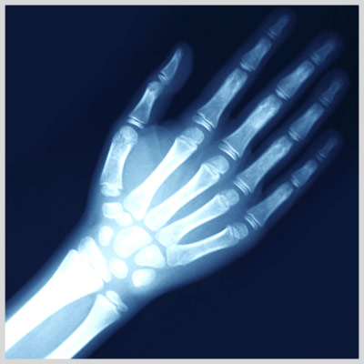
X-Ray Fluoroscopy
One of the earliest applications of x-rays was in medicine, where they were used for both diagnosis and therapy. They penetrate soft tissues but are stopped by bones, which absorb them. Thus if a photographic plate that is sensitive to x-rays is placed behind a part of the body and an x-ray source is placed in front, x-ray exposure will result in an image of the bones and internal organs. When the radiograph, or plate, is developed, a negative image is produced. Click here for more details.
Click Here to Read more
Since their discovery in 1895, x-rays have been a vital scientific tool, revealing previously concealed worlds. Because of their great penetrating power, x-rays can also be used to study the structure of living organisms.
One of the earliest applications of x-rays was in medicine, where they were used for both diagnosis and therapy. They penetrate soft tissues but are stopped by bones, which absorb them. Thus if a photographic plate that is sensitive to x-rays is placed behind a part of the body and an x-ray source is placed in front, x-ray exposure will result in an image of the bones and internal organs. When the radiograph, or plate, is developed, a negative image is produced. Tissues that are easily penetrated by x-rays appear dark, while bones and dense tissues show up as light or white regions.
Although bones are the most opaque structures, there are many dense tissues, such as tumors, that can also show up unusually light in radiographs. These images can be used to study damaged or broken bones, inspect dental cavities, detect foreign objects in the body, and diagnose diseases.
To utilize x-rays for the investigation of other, less dense tissues of the body, such as the gastrointestinal tract, the tissues must first be made opaque to x-rays. Generally, patients are asked to drink a mixture containing an opaque substance, such as barium, so that the outline of the digestive tract becomes visible with x-rays.
The clinic also offers fluoroscopy, a similar technique that uses x-rays to observe a particular organ in action, such as the heart. The patient is placed in the fluoroscope between the x-ray source and a screen that is coated with a fluorescent substance. The image that appears when the x-rays strike the screen is brightened by an electronic device called an image intensifier.


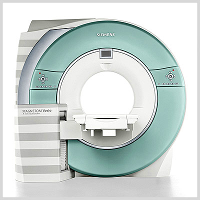
3T MRI
Introducing our brand new state of the art Simens Magnetom Verio 3T, featuring 70-cm open-style bore. The Verio is designed to accommodate large patients, as well as ease fears from claustrophobic patients. The Verio offers a new software program that will allow for faster exams and improved patient positioning. Advanced imaging center is the first to offer this technology within a 70 mile radius. Click here for more details.
Click Here to Read more
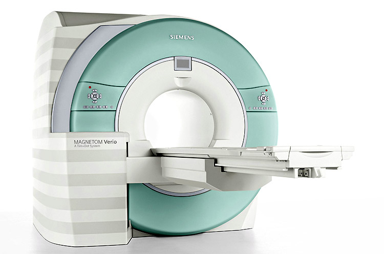
Introducing our brand new state of the art Simens Magnetom Verio 3T, featuring 70-cm open-style bore. The Verio is designed to accommodate large patients, as well as ease fears from claustrophobic patients. The Verio offers a new software program that will allow for faster exams and improved patient positioning. Advanced imaging center is the first to offer this technology within a 70 mile radius.
3T MRI
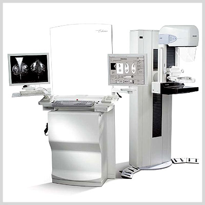
Announcing our new Hologic Selenia mammogram with CAD (Computer-Aided Detection). Our new mammogram will be able to indentify region of interest and bring them to the attention of the radiologist, helping to decrease false negative reading. The Hologic Selenia features a selenium based direct capture technology that eliminate light diffusion completely, providing perfect clarity and exquisite image quality. Our Hologic Selenia mammogram can help detect cancer up to 2 years before it can be found by a physical examination. Click here for more details.
Click Here to Read more
Announcing our new Hologic Selenia mammogram with CAD (Computer-Aided Detection). Our new mammogram will be able to indentify region of interest and bring them to the attention of the radiologist, helping to decrease false negative reading. The Hologic Selenia features a selenium based direct capture technology that eliminate light diffusion completely, providing perfect clarity and exquisite image quality. Our Hologic Selenia mammogram can help detect cancer up to 2 years before it can be found by a physical examination.
ImageChecker CAD
As a pioneer that delivered the industry's most widely used CAD solutions, we are confident that we have the right CAD solution for your practice. The majority of radiologists in the U.S. use CAD to review mammography studies in order to identify suspicious lesions and help reduce false negatives.
ImageChecker CAD for Digital Mammography
ImageChecker CAD can process images from most direct capture full-field digital mammography (FFDM) detectors and displays them on a range of workstation environments. The display of digital CAD marks depends upon the viewing solution chosen. Whichever display you choose, basic RightOn CAD marks will appear on all displays. However, the SecurView DX workstation is the only softcopy diagnostic workstation that can display the most advanced CAD capabilities.
OUR LOCATIONS


